Breast cancer is the most commonly diagnosed cancer among American women with about one in eight women expected to develop the disease during their lifetime. However, there is good news that comes along with this statistic.
The five-year relative survival rate for localized breast cancer—disease that has not spread outside the breast—is 99%. And the survival rate is 86% in cases where the cancer has only spread outside the breast to nearby lymph nodes.1
The key to these success stories is early detection and treatment which are both possible due to major advances in breast cancer imaging. This article offers an overview of the different types of breast cancer imaging services that are available to women and how they are used to make a diagnosis.
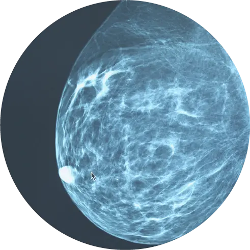
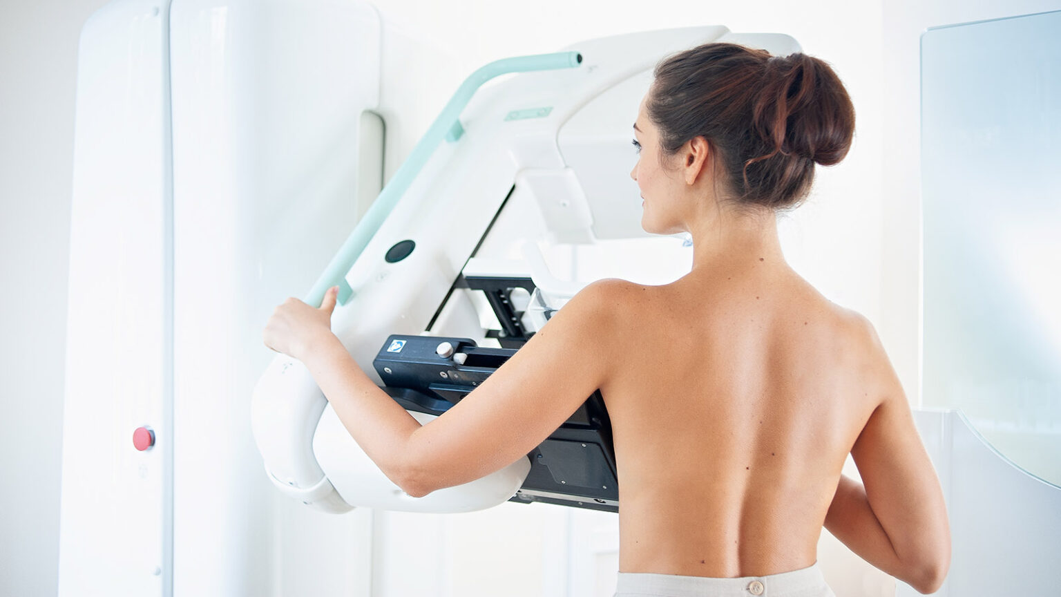
Screening Mammogram
A screening mammogram uses low-dose X-rays to take images of each breast. These exams can detect cancer months and even years before symptoms emerge.
During a screening mammogram, you will stand in front of the X-ray machine and then a trained technologist will place your breast on a plastic plate. An additional plate will firmly press down on your breast to hold it in place while the imaging takes place.2 You may feel some breast pressure during the imaging, but it shouldn’t hurt. The entire exam takes only about 20 minutes.3
The general guidelines are for women of average risk to begin annual screening mammograms at the age of 40.4
Gradually replacing traditional mammography, 3D mammograms are helpful for making a diagnosis when women have dense breast tissue. A 3D mammogram produces the same amount of low-dose radiation and the cost of the procedure is also covered by insurance.
Also known as digital breast tomosynthesis, the procedure is similar to traditional mammography, except the X-ray equipment takes multiple images and combines them to provide a 3D view.7
You will receive a report of your imaging procedure within about two weeks. If the mammogram reveals something abnormal or if the images are unclear, you may need follow-up tests.
Diagnostic Mammogram
A diagnostic mammogram is used to follow up on any abnormal results found in a screening mammogram. This test is similar to a screening mammogram, but it involves taking more images and more detailed pictures of the breast.
A diagnostic mammogram also is used when one or more symptoms are present. These can include:
- a lump in the breast
- breast pain
- changes in breast size or shape
- nipple discharge
- thickening of breast skin
Diagnostic mammograms are usually longer than traditional mammograms, provide more views of your breast, and zoom in on more specific areas compared to screening mammograms.5
Breast Ultrasound
A breast ultrasound is another tool doctors use to follow up on any suspicious findings from a clinical exam or a mammogram.
The tests use high-frequency sound waves to take images of the inside of the breast. The sound waves bounce off firm breast tissue to reveal any irregularities. For example, they can indicate the difference between a cyst that is filled with liquid and a solid mass.
Your doctor may recommend a beast ultrasound for one of the following reasons:
- You have dense breast tissue that makes clear mammogram imaging challenging
- You are pregnant (An ultrasound does not use radiation, making it safer for your unborn child)
- You are age 25 or younger6
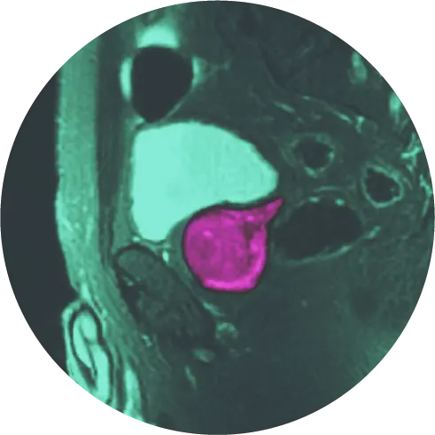
Breast MRI
A breast MRI (magnetic resonance imaging) test uses magnetic fields to create detailed images of the breast. During a breast MRI, fluids are injected into the breast through an IV in order to improve the visibility of the inside of the breast.
Your doctor may recommend this test as a follow-up to other test results in order to make a precise diagnosis. It is typically used to screen women who have a higher-than-average risk of getting breast cancer.8
Schedule a Consultation with Breast Cancer Imaging Experts
With early detection and the successful treatment options that are available today, the future of breast care is promising and bright.
If you have additional questions about breast cancer screenings or would like to schedule an appointment, please give our experts at HALO Precision Diagnostics™ a call at 877-225-2831 or fill out the form below.
References
- https://www.womenshealth.gov/blog/99-percent-survival-rate-breast-cancer-caught-early
- https://www.komen.org/breast-cancer/screening/
- https://www.cdc.gov/cancer/breast/basic_info/mammograms.htm#:~:text=You%20will%20stand%20in%20front,X%2Dray%20is%20being%20taken.
- https://www.jacr.org/article/S1546-1440(17)31524-7/fulltext
- https://www.nationalbreastcancer.org/diagnostic-mammogram
- https://www.hopkinsmedicine.org/health/treatment-tests-and-therapies/breast-ultrasound
- https://www.mdanderson.org/publications/focused-on-health/FOH-3D-mammography.h19-1589835.html
- https://www.cancer.org/cancer/breast-cancer/screening-tests-and-early-detection/breast-mri-scans.html